Biology
Biology Lab: Microscope Basic Skills

Lab Exercise
Biology Lab: Microscope Use
Goal: Learn to use a microscope and calibrate its field of view for different objectives.
Read about microscopes and their accessories in Illustrated Guide to Home Biology Experiments pp. 10-32. Be sure that you can identify the parts and functions of a compound microscope.
Read through Lab I-1 and perform Procedure I-1-1. If you have time, experiment with the Darkfield Apparatus lighting methods and compare details on a low-magnification object.
Use the BioNetwork Virtual Microscope to become acquainted with the use of a binocular microscope. Read throug the short guide, the work through the "Learn" module, clicking on different objects to discover their use. Then use the Explore option to look at at least two different slides adjusting light, focus, and magnification. How do you adjust coarse focus? Fine focus? Lighting?
Alternate lab: microscope tour
Read the procedure below, which we used prior to the publication of the IGHBE. You do not need to repeat the activities, but it's worth checking out variations in terminology and methods for calibrating the field of view and magnification levels.
Materials
- Microscope*
- Hand lens (2 if possible)
- Clean blank microscope slides
- Ruler (marked in mm if possible)
- Fibers like hair, cotton thread, silk
- Granular material: small amounts of salt, sugar, cornstarch, baking soda, epsom salts
- Pencil, plain and/or graph paper for drawing
Procedure
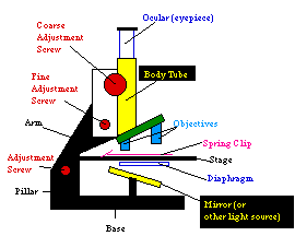
Study the rules in the instruction manual that came with your microscope for its use and care. The procedure below will work most of the time, but if your manual says otherwise, follow it instead. For each observation you make, record the magnification and diaphragm stop used. You can calculate the total magnification by multiplying the eyepiece magnification times the objective magnification. For example, if the eyepiece is 10X and the objective is 10X, the total magnification is 10X * 10X = 100X.
- Practice positioning and focusing your microscope. Use a prepared slide, or a hair or thread.
- Use the coarse adjustment to raise the tube high.
- Tilt the arm until you can look through the eyepiece comfortably. You may have to adjust the arm screw so that it will tilt; remember to tighten it so the arm will stay in position.
- If you have a diaphragm, adjust it so that you are using the largest hole.
- Adjust the mirror or light source so that you have the maximum light.
- Set the objectives so you are using the one with lowest magnification.
- Put your slide in place on the stage, and center the specimen over the light hole. Fasten in place with the spring clips.
- Using the coarse adjustment, and looking at the microscope from the side, lower the tube until it almost touches the slide. Do not mash the objective into your specimen!
- Now, looking through the eyepiece, raise the objective with the coarse adjustment until you see your specimen.
- Use the fine adjustment to bring your specimen into focus.
- Compare magnifications. Use a single specimen, which should be translucent (light-passing, such as onion skin), if possible, for the following exercise.
- Observe the specimen at the lowest eyepiece and objective magnification. Note the eyepiece and objective magnifications and calculate the total magnification. Make a rough drawing showing the size of the specimen in relation to the field of view (the circle you see through the eyepiece).
- Change the objective or eyepiece to a higher magnification. What do you notice about the size of your specimen? What do notice about the amount of light?
- Compare light amounts. Change the amount of light coming through the eyepiece, either by changing your light source or by setting the diaphragm to a smaller opening. How does changing the light change your ability to see an object? Is more light always better? When should you use less light?
- Determine orientation. Use a specimen with a definite orientation (differently shaped ends?).
- Place the slide on the microscope stage and notice the orientation of the specimen, then look at it through the eyepiece. How does it look? Has the microscope reversed up and down? Left and right?
- While looking through the microscope, move the slide slightly to the left. Which way does your specimen move?
- Practice moving the specimen around until you are comfortable centering it.
- Determine the field of view. Reset the microscope to the lowest power, and look at your ruler through it. Estimate the diameter of the field of view, measure from the center of the marks on the ruler. Make a notation of the size of the field in mm or fractions of an inch, and the magnification you are using.
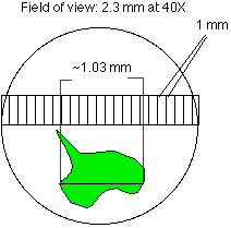
- You can calculate the size of the field of view at other magnifications. Multiply your field of view at the low magnification by the ratio of the low magnification to the higher one. If you measures a field 2.5mm across at 40X, then the field of view at 200X will be 2.5 X (40/200) = .5mm.
- Resolving power. The resolving power of a magnification system is its ability to help you differentiate between two close objects. Your eye will see as one thing objects which are too close together. A microscope or telescope will show the objects separated by some distance.
- Now that you have some experience with your microscope, look at some of the following:
- Salt, sugar, corn starch, baking soda, flour, epsom salts. What problems do you have focusing crystal solids?
- Hair, cotton thread, nylon thread, silk thread.
- Put a drop of water on the slide and put a grain of salt or sugar on it, and watch the grain dissolve.
- Put a drop of water on the slide and with the end of a pin, add a droplet of vegetable oil. Look at the edges of the drop.
- Pond water, soil samples, spider silk from your survey area. (We'll do water animals some more when we get to protista).
- Have fun!
*If you do not have access to a microscope, try the following:
- Position a light source so that it will fall on your slide from above, or from below, but not shine directly upward through your lens system.
- Fix a piece of glass (a microscope slide if you have one) so the light shines on or through it.
- Fix one hand lens (if you have two, use the one with greater magnification) in position so you can look down at your "slide". Once the mirror, slide and first lens are in place, you should not have to move them.
- Use the second lens to look through the first at your slide. You have just jury-rigged a microscope! It probably won't have the power of a commercial one, and you will have to do more work to look at your sample, but you will still be able to do much of the lab, including estimating the "power" of your lenses.
Report Format
Your lab report should include
- a description of your equipment
- each procedure step, especially if you improvised or improved upon the suggested procedure;
- answers to all the questions asked in the procedure (this includes your calculations for the field of view and the size of one specimen viewed at two different magnifications)
- descriptions (including size estimates) of at least two items you examined.
© 2005 - 2024 This course is offered through Scholars Online, a non-profit organization supporting classical Christian education through online courses. Permission to copy course content (lessons and labs) for personal study is granted to students currently or formerly enrolled in the course through Scholars Online. Reproduction for any other purpose, without the express written consent of the author, is prohibited.